Not all foreign bodies embedded in the arm or leg need removal. There
are many veterans of war, industrial accident, and other misfortunes who
have such objects, often metallic, which give rise to no symptoms and
threaten no complications. For those that do however need operative removal,
this may be a vexing and sometimes unsuccessful venture.
To minimize the operating time and the likelihood of failure in locating a foreign body in a limb, there are several steps it is important not to omit. First, a careful history may indicate the site, nature, and size of the foreign body. Examining tin object from which it may have broken off may be helpful. Inspection will show the puncture wound of entrance if it is recent, or a scar. Palpation may indicate the object if it is superficial.
X-rays should be taken in two planes, with a radio-opaque marker consisting of two crossed wires (for which pieces of paper clip may serve) fastened to the skin with cellotape. Their intersection should be sited exactly at the puncture wound or over an indelible ink mark. Sometimes an object can be felt unmistakably just under the skin, and it is permissible then to make an incision over it under local anaesthesia. But for any object not clearly palpable this is the way to frustration and failure.
Formal exploration for a foreign body in the tissues of a limb requires general or regional anaesthesia, a bloodless field, and a trained operator; it is not something to be tackled by the inexperienced or the unpractised.
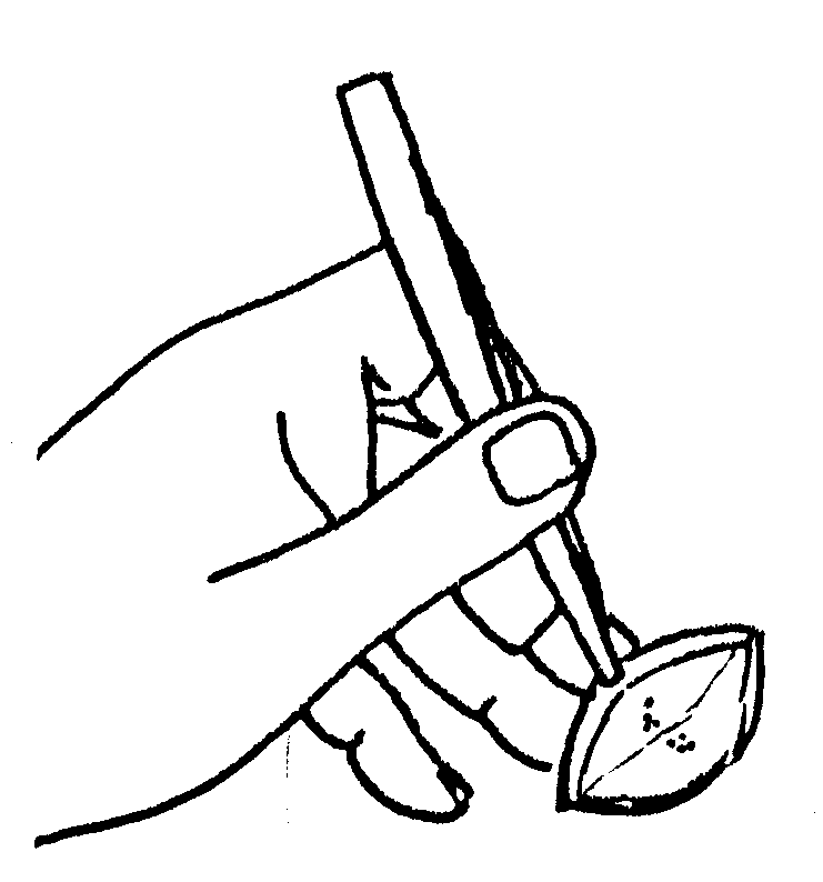 Fig. 1. Elevating the tissue flap
[Note the double hand function, with a precision grip for the forceps, and the fuilcrum of ring fingertip to stretch the skin over]
The track of penetration, if recent, is marked by a little persisting haemorrhage and bruising which can be seen only in a bloodless field (see figure); normal capillary ooze - or worse - would completely obscure these signposts to the object being sought.
With the Xrays on a viewing box in the operating theatre, the initial incision should create a flap so constructed that it must contain or overlie the foreign body. This is in preference to a straight incision. The flap is deepened to deep fascia and then raised.Sometimes there is a little sterile pus about the foreign body, especially if it has been present for some weeks. This may be cultured, but there is rarely further trouble, and the wound does not need to be drained. The wound is sutured with interrupted silk or nylon stitches to the skin.
-o0o-
Removing foreign bodies from the limbs
Michael Patkin and Gordon Kerridge Australian Family Physician, Vol. 1,
April / May, 1972 138
_____________
Michael Patkin, MB, BS, FRCS(Eng), FRCS(Ed), FRACS, is a Fellow in Surgery at Royal Newcastle Hospital and Gordon Kerridge, MB, BS, FRCS(Eng), FRCS(Ed), FRACS, FACS, is Director' of Orthopaedics and Traumatic Surgery, Royal Newcastle Hospital.
Reprint requests
Dr. G. Kerridge, Director of Orthonuedics and Traumatic Surgery, Royal
Newcastle Hospital, Newcastle, N.S.W. 2300.
________
When dealing with foreign bodies surgically, don't forget about tetanus, gas gangrene, and lawyers!